VC. Voronov Nuclear magnetic resonance
NUCLEAR MAGNETIC RESONANCE(NMR), the phenomenon of resonant absorption of radio frequency electromagnet. energy in-tion with non-zero magn. moments of nuclei located in ext. permanent mage. field. Non-zero nuclear magnet. 1 H, 2 H, 13 C, 14 N, 15 N, 19 F, 29 Si, 31 P, and other nuclei have momentum. NMR is usually observed in a uniform constant magn. field B 0 ,
a weak radio-frequency field B 1 perpendicular to the field B 0 is superimposed on a cut. For in-in, in which the nuclear spin I \u003d 1/2 (1 H, 13 C, 15 N, 19 F, 29 Si, 31 P, etc.), in the field B 0, two orientations of the magnet are possible. dipole moment of the nucleus "along the field" and "against the field". The emerging two levels of energy E due to the interaction. magn. momentum of the nucleus with field B 0
separated by an interval
Provided that or where h is Planck's constant, v 0 is the frequency of the radio frequency field B 1, is the circular frequency, - the so-called. gyromag. ratio of the nucleus, resonant absorption of the energy of the field B 1 ,
called NMR. For nuclides 1 H, 13 C, 31 P, the NMR frequencies in the field B 0 = 11.7 T are equal, respectively. (in MHz): 500, 160.42 and 202.4; values (in MHz/T): 42.58, 10.68 and 17.24. According to the quantum model, 2I + 1 energy levels arise in the field B 0, transitions between which are allowed at where m is the magn. quantum number.
Experiment technique. Parameters of the NMR spectra. NMR spectroscopy is based on the NMR phenomenon. NMR spectra are recorded using radio spectrometers (Fig.). A sample of the investigated in-va is placed as a core in the coil of the generating circuit (field B 1), located in the gap of the magnet, which creates the field B 0 so that when resonant absorption occurs, which causes a voltage drop in the circuit, the circuit of which includes a coil with sample. The voltage drop is detected, amplified, and fed to an oscilloscope sweep or recording device. In modern Radio NMR spectrometers usually use magician, field strength of 1-12 T. The region of the spectrum, in which there is a detectable signal with one or more. maxima, called NMR absorption line. Width of the observed line, measured at half max. intensity and expressed in Hz, called. NMR line width. Resolution of the NMR spectrum - min. width of the NMR line, to-ruyu allows you to observe this spectrometer. Transmission speed - speed (in Hz / s), with which the magnetic strength changes. field or frequency of RF radiation affecting the sample when obtaining the NMR spectrum.

Scheme of the NMR spectrometer: 1 - coil with the sample; 2 - magnet poles; 3 - radio frequency field generator; 4 - amplifier and detector; 5 - modulating voltage generator; 6 - field modulation coils B 0; 7 - oscilloscope.
The system redistributes the absorbed energy within itself (the so-called spin-spin, or transverse relaxation; characteristic time T 2) and gives it to the environment (spin-lattice relaxation, relaxation time T 1). Times T 1 and T 2 carry information about internuclear distances and correlation times decomp. they say movements. Measurements of the dependence of T 1 and T 2 on t-ry and frequency v 0 provide information on the nature of thermal motion, chemical. equilibria, phase transitions, etc. In solids with a rigid lattice, T 2 \u003d 10 μs, and T 1\u003e 10 3 s, since there is no regular mechanism of spin-lattice relaxation and relaxation is due to paramagnetism. impurities. Due to the smallness of T 2 , the natural width of the NMR line is very large (tens of kHz), and their registration is in the NMR region of broad lines. In liquids of low viscosity, T 1 T 2 is measured in seconds. Resp. NMR lines have a width of the order of 10 -1 Hz (high-resolution NMR). For an undistorted reproduction of the line shape, it is necessary to pass through a line with a width of 0.1 Hz for 100 s. This imposes significant limitations on the sensitivity of NMR spectrometers.
The main parameter of the NMR spectrum is chem. shift - taken with the appropriate sign, the ratio of the difference between the frequencies of the observed NMR signal and some conventionally selected reference signal to.-l. standard to the frequency of the reference signal (expressed in parts per million, ppm). Chem. NMR shifts are measured in dimensionless values counted from the peak of the reference signal. If the standard gives a signal at a frequency v 0, then ![]() Depending on the nature of the nuclei under study, proton NMR, or PMR, and 13 C NMR are distinguished (tables of chemical shifts are given on the flyleaves of the volume). NMR 19 F (see Organofluorine compounds), NMR 31 P (see Organophosphorus compounds), etc. The values have a significant characteristic and make it possible to determine the presence of certain mol from NMR spectra. fragments. Relevant data on chem. shifts diff. kernels are published in reference and manuals, and also are brought in databases, to-rymi are supplied with modern. NMR spectrometers. In the series of compounds close in structure, chemical. the shift is directly proportional to the electron density on the corresponding nuclei.
Depending on the nature of the nuclei under study, proton NMR, or PMR, and 13 C NMR are distinguished (tables of chemical shifts are given on the flyleaves of the volume). NMR 19 F (see Organofluorine compounds), NMR 31 P (see Organophosphorus compounds), etc. The values have a significant characteristic and make it possible to determine the presence of certain mol from NMR spectra. fragments. Relevant data on chem. shifts diff. kernels are published in reference and manuals, and also are brought in databases, to-rymi are supplied with modern. NMR spectrometers. In the series of compounds close in structure, chemical. the shift is directly proportional to the electron density on the corresponding nuclei.
The generally accepted standard for NMR and 13 C NMR is tetramethylsilane (TMS). Standard m. b. dissolved in the investigated solution (internal standard) or placed, for example, in a sealed capillary located inside the sample ampoule (external standard). As solvents, only those whose own absorption does not overlap with the region of interest for the study can be used. For PMR, the best p-carriers are those that do not contain protons (CC1 4 , CDC1 3 , CS 2 , D 2 O, etc.).
In polyatomic molecules, the nuclei of identical atoms occupying chemically non-equivalent positions have different chemical. shifts due to the difference in magnetic. screening of nuclei by valence electrons (such nuclei are called anisochronous). For the i-th core ![]() where is the constant diamagnet. screening, measured in ppm. For protons, the typical range of changes is up to 20 ppm; for heavier nuclei, these intervals are 2-3 orders of magnitude larger.
where is the constant diamagnet. screening, measured in ppm. For protons, the typical range of changes is up to 20 ppm; for heavier nuclei, these intervals are 2-3 orders of magnitude larger.
An important parameter of the NMR spectra is the constant of the spin-spin interaction. (constant CER) - a measure of indirect CER between dec. magn. nuclei of one molecule (see Spin-spin interaction); expressed in Hz.
Intermod. nuclear spins with electron spins contained in the molecule between the nuclei i and j, lead to the mutual orientation of these nuclei in the field B 0 (CNE). With sufficient resolution ![]() CER leads to additional. multiplicity of lines corresponding to certain values of chemical. shifts: where J ij - CER constants; F ij - quantities, the values of which are determined by the spins of the nuclei i and j, the symmetry of the corresponding pier. fragment, dihedral angles between the chemical. bonds and the number of these bonds between the nuclei participating in the CER.
CER leads to additional. multiplicity of lines corresponding to certain values of chemical. shifts: where J ij - CER constants; F ij - quantities, the values of which are determined by the spins of the nuclei i and j, the symmetry of the corresponding pier. fragment, dihedral angles between the chemical. bonds and the number of these bonds between the nuclei participating in the CER.
If chem. If the shifts are large enough, i.e. min max (J ij), then the SWV appear as simple multiplets with a binomial distribution of intensities (first-order spectra). Thus, in the ethyl group, the methyl proton signal appears as a triplet with an intensity ratio of 1:2:1, and the methylene proton signal appears as a quadruplet with an intensity ratio of 1:3:3:1. In the 13 C NMR spectra, methine groups are doublets (1:1), and methylene and methyl groups are respectively. triplets and quadruplets, but with higher than in the proton spectrum, the values of the CER constants. Chem. the shifts in the spectra of the first order are equal to the intervals between the centers of the multiplets, and J ij - to the distances between adjacent peaks of the multiplet. If the first order condition is not satisfied, then the spectra become complex: in them, not a single interval, generally speaking, is equal to either J ij . The exact values of the spectral parameters are obtained from quantum mech. calculations. Relevant programs are included in the mat. providing modern NMR spectrometers. Informativeness of chem. shifts and CCR constants has turned high-resolution NMR spectroscopy into one of the most important quality methods. and quantities. analysis of complex mixtures, systems, preparations and compositions, as well as studies of the structure and reactions. the ability of molecules. When studying conformations, degenerate and other dynamic. systems, geom. structures of protein molecules in solution, with non-destructive local chem. analysis of living organisms, etc. the possibilities of NMR methods are unique.
Nuclear magnetization in-va. In accordance with the Boltzmann distribution in a two-level spin system of N spins, the ratio of the number of spins N + at the lower level to the number of spins N - at the upper level is where k is Boltzmann's constant; T - t-ra. At B 0 \u003d 1 T and T \u003d 300 K for protons, the ratio N + /N - .= 1.00005. This ratio determines the magnitude of the nuclear magnetization in-va, placed in the field B 0 . Magn. moment m each nucleus performs a precessional motion about the z-axis, along which the field B 0 is directed; the frequency of this movement is equal to the NMR frequency. The sum of the projections of precessing nuclear moments on the z axis forms a macroscopic. magnetization in-va ![]() M z \u003d 10 18 In the xy plane, perpendicular to the z axis, the projections of the vectors are equal to zero due to the randomness of the precession phases: M xy \u003d 0. Energy absorption during NMR means that more spins pass from the lower level to the upper one per unit time than in in the opposite direction, i.e., the population difference N + - N - decreases (heating of the spin system, NMR saturation). When saturated in the stationary regime, the magnetization of the system can greatly increase. This is the so-called. the Overhauser effect, for nuclei designated NOE (Nuclear Overhauser effect), to-ry is widely used to increase sensitivity, as well as to assess internuclear distances in the study of mol. geometries by NMR spectroscopy methods.
M z \u003d 10 18 In the xy plane, perpendicular to the z axis, the projections of the vectors are equal to zero due to the randomness of the precession phases: M xy \u003d 0. Energy absorption during NMR means that more spins pass from the lower level to the upper one per unit time than in in the opposite direction, i.e., the population difference N + - N - decreases (heating of the spin system, NMR saturation). When saturated in the stationary regime, the magnetization of the system can greatly increase. This is the so-called. the Overhauser effect, for nuclei designated NOE (Nuclear Overhauser effect), to-ry is widely used to increase sensitivity, as well as to assess internuclear distances in the study of mol. geometries by NMR spectroscopy methods.
Vector NMR model. When registering NMR, a radio frequency field is applied to the sample, acting in the xy plane. In this plane, the field B 1 can be considered as two vectors with amplitudes B 1m/ 2, rotating with frequency in opposite directions. A rotating coordinate system x "y" z is introduced, the x-axis coincides with the vector B 1m / 2, rotating in the same direction as the vectors. Its action causes a change in the angle at the top of the cone of precession of nuclear magnetic moments; nuclear magnetization M z begins to depend on time, and a non-zero projection of nuclear magnetization appears in the x "y" plane. In a fixed coordinate system, this projection rotates with frequency, i.e., a radio frequency voltage is induced in the inductor, which, after detection, gives the NMR signal - the function of nuclear magnetization from frequency is distinguished by a slow change (sweep mode) and pulsed NMR.The real complex movement of the nuclear magnetization vector creates two independent signals in the x "y" plane: M x, (in-phase with radio frequency voltage B 1) and M y" (shifted relative to B 1 in phase by 90 ° C). Simultaneous registration of M x" and M y" (quadrature detection) doubles the sensitivity of the NMR spectrometer. With a sufficiently large amplitude B 1m of the projection M z = M x" = M y" = 0 (NMR saturation). Therefore, with the continuous action of the field B 1, its amplitude must be very small in order to keep the initial conditions of observation unchanged.
In pulsed NMR, the value of B 1 , on the contrary, is chosen so large that during the time t and T 2 the vector M z deviates from the z axis by an angle . At \u003d 90 °, the impulse is called 90 ° (/2-impulse); under its influence, the nuclear magnetization vector turns out to be in the x "y" plane, i.e., after the end of the pulse, the vector M y "begins to decrease in amplitude with time T 2 due to the phase divergence of its constituent elementary vectors (spin-spin relaxation). Restoration of the equilibrium of nuclear magnetization M z occurs with the spin-lattice relaxation time T 1. At = 180° (momentum), the vector M z fits along the negative direction of the z axis, relaxing after the end of the pulse to its equilibrium position. Combinations and pulses are widely used in modern multi-pulse versions NMR spectroscopy.
An important feature of the rotating coordinate system is the difference between the resonant frequencies in it and in the fixed coordinate system: if B 1 B lok (static local field), then the vector M precesses in the rotating coordinate system relative to the field. When finely tuned to resonance, the NMR frequency in the rotating coordinate system This makes it possible to significantly expand the possibilities of NMR in the study of slow processes in matter.
Chem. exchange and NMR spectra(dynamic. NMR). The parameters of the two-position exchange A B are the sojourn times and as well as the sojourn probabilities ![]() and At low t-re, the NMR spectrum consists of two narrow lines separated by Hz; then, with decreasing and, the lines begin to broaden, remaining in their places. When the exchange frequency begins to exceed the initial distance between the lines, the lines begin to converge, and at a 10-fold excess, one wide line is formed in the center of the interval (v A, v B), if With a further increase in temperature, this combined line becomes narrow. Comparison of experiments. spectrum with the calculated allows for each t-ry to specify the exact frequency of chemical. exchange, these data calculate thermodynamic. process characteristics. With multipositional exchange in a complex NMR spectrum, theoret. the spectrum is obtained from quantum mech. calculation. Dynamic NMR is one of the main methods for studying stereochemistry. non-rigidity, conformational equilibria, etc.
and At low t-re, the NMR spectrum consists of two narrow lines separated by Hz; then, with decreasing and, the lines begin to broaden, remaining in their places. When the exchange frequency begins to exceed the initial distance between the lines, the lines begin to converge, and at a 10-fold excess, one wide line is formed in the center of the interval (v A, v B), if With a further increase in temperature, this combined line becomes narrow. Comparison of experiments. spectrum with the calculated allows for each t-ry to specify the exact frequency of chemical. exchange, these data calculate thermodynamic. process characteristics. With multipositional exchange in a complex NMR spectrum, theoret. the spectrum is obtained from quantum mech. calculation. Dynamic NMR is one of the main methods for studying stereochemistry. non-rigidity, conformational equilibria, etc.
Rotation at a magical angle. Expression for the potential of the dipole-dipole interaction. contains multipliers ![]() where is the angle between B 0 and the internuclear vector r ij . At = arccos 3 -1/2 = 54 ° 44 "("magic" angle), these factors vanish, i.e., the corresponding contributions to the line width disappear. If a solid sample is twisted at a very high speed around an axis inclined under the magic angle to B 0 , then high-resolution spectra with almost as narrow lines as in a liquid can be obtained in a solid.
where is the angle between B 0 and the internuclear vector r ij . At = arccos 3 -1/2 = 54 ° 44 "("magic" angle), these factors vanish, i.e., the corresponding contributions to the line width disappear. If a solid sample is twisted at a very high speed around an axis inclined under the magic angle to B 0 , then high-resolution spectra with almost as narrow lines as in a liquid can be obtained in a solid.
Broad lines in solids. In crystals with a rigid lattice, the shape of the NMR line is due to static. distribution of local magnets. fields. All lattice nuclei, with the exception of the cluster , in the translation-invariant volume V 0 around the considered nucleus, give a Gaussian distribution g(v) = exp(-v 2 /2a 2), where v is the distance from the center of the line; the width of the Gaussian a is inversely proportional to the mean geom. volumes V 0 and V 1, and V 1 characterizes the average concentration of magnesium throughout the crystal. nuclei. Inside V 0 concentration of magnetic. nuclei more than average, and near nuclei due to the dipole-dipole interaction. and chem. shifts create a spectrum limited to the interval (-b, b), where b is approximately twice as large as a. In the first approximation, the spectrum
1.1. From the history of magnetic resonance spectroscopy.
Until recently, our ideas about the structure of atoms and molecules were based on studies using optical spectroscopy methods. In connection with the improvement of spectral methods, which have advanced the field of spectroscopic measurements into the range of ultrahigh (approximately 10 3 - 10 6 MHz; microradio waves) and high frequencies (approximately 10 -2 - 10 2 MHz; radio waves), new sources of information about the structure of matter have appeared. During the absorption and emission of radiation in this frequency range, the same basic process occurs as in other ranges of the electromagnetic spectrum, namely, when moving from one energy level to another, the system absorbs or emits a quantum of energy.
The energy difference between the levels and the energy of the quanta participating in these processes are about 10 -7 eV for the radio frequency region and about 10 -4 eV for microwave frequencies.
The existence of nuclear moments was first discovered when studying the hyperfine structure of the electronic spectra of some atoms using high-resolution optical spectrometers.
The hyperfine structure of atomic spectra led Pauli in 1924 to the idea that some nuclei have a moment of momentum (angular momentum) and, consequently, a magnetic moment that interacts with atomic orbital electrons. Subsequently, this hypothesis was confirmed by spectroscopic measurements, which made it possible to determine the values of the angular and magnetic moments for many nuclei.
Under the influence of an external magnetic field, the magnetic moments of the nuclei are oriented in a certain way, and it becomes possible to observe transitions between nuclear energy levels associated with these different orientations: transitions that occur under the action of radiation of a certain frequency. The quantization of the energy levels of the nucleus is a direct consequence of the quantum nature of the angular momentum of the nucleus receiving 2 I+ 1 values. Spin quantum number (spin) I can take any value multiple of 1/2; 176 71 Lu has the highest known I value (≥7). The measurable largest value of the angular momentum (the largest value of the projection of the moment on the selected direction) is equal to Iħ, where ħ=h/2π, and h is Planck's constant.
Values I it is impossible to predict for specific nuclei, but it has been observed that isotopes in which both mass number and atomic number are even have I= 0, and isotopes with odd mass numbers have half-integer spins. Such a situation, when the numbers of protons and neutrons in the nucleus are even and equal ( I= 0) can be considered as a state with "complete pairing", similar to the complete pairing of electrons in a diamagnetic molecule.
In 1921 Stern and Gerlach showed by the atomic beam method that the measurable values of the magnetic moment of an atom are discrete, corresponding to the spatial quantization of an atom in an inhomogeneous magnetic field. In subsequent experiments, passing a beam of hydrogen molecules through a constant magnetic field, it was possible to measure the small magnetic moment of the hydrogen nucleus. Further development of the method consisted in the fact that the beam was affected by an additional magnetic field oscillating at a frequency at which transitions between nuclear energy levels corresponding to the quantum values of the nuclear magnetic moment are induced.
If the nuclear spin number is I, then the nucleus has (2I+1) equally spaced energy levels; in a constant magnetic field with strength H, the distance between the highest and lowest of these levels is 2mH, where m is the maximum measurable value of the magnetic moment of the nucleus. Hence, the distance between adjacent levels is equal to mH/I, and the frequency of the oscillating magnetic field, which can cause transitions between these levels, is equal to mH/Ih.
In an experiment with a molecular beam, those molecules reach the detector whose energy does not change. The frequency at which resonant transitions between levels occur is determined by successively changing (sweeping) the frequency in a certain range. At a certain frequency, there is a sudden decrease in the number of molecules reaching the detector.
The first successful NMR observations of this kind were made with fundamental magnetic fields of the order of several kilooersteds, which correspond to frequencies of an oscillating magnetic field in the range of 10 5 -10 8 Hz. Resonance energy exchange can occur not only in molecular beams; it can be observed in all aggregate states of matter.
In 1936 Horner tried to discover the resonance of Li 7 nuclei in lithium fluoride and H 1 nuclei in potassium alum. Another unsuccessful attempt was made by Gortner and Brur in 1942. Registration of the absorption of high-frequency energy at resonance in these experiments was supposed to be carried out, respectively, by the calorimetric method and by anomalous dispersion. The main reason for the failure of these experiments was the choice of unsuitable objects. Only at the end of 1945, two groups of American physicists led by F. Bloch and E.M. Purcell were the first to receive nuclear magnetic resonance signals. Bloch observed resonant absorption by protons in water, and Purcell was successful in discovering nuclear resonance by protons in paraffin. For this discovery, they were awarded the Nobel Prize in 1952.
1.2. Technological applications of NMR (the main advantages of the NMR method).
The NMR method, although it is called the method of nuclear magnetic resonance, has nothing to do with nuclear physics, which, as is known, studies the processes of nuclear transformation, i.e. radioactive processes. In this case, the magnetic energy (and the NMR phenomenon takes place when the sample under study is placed in a constant magnetic field) does not affects on the thermodynamic properties of matter, because it is many times (more precisely, several orders of magnitude) less than the thermal energy characteristic of processes occurring under normal conditions, including biological ones.
The main advantages of the NMR method.
- High resolution - on ten orders more than optical spectroscopy.
Ability to lead quantitative accounting (counting) of resonating nuclei. This opens up opportunities for quantitative analysis of the substance.
The NMR spectra depend on the nature of the processes occurring in the substance under study. Therefore, these processes can be studied by the indicated method. Moreover, it is available temporary the scale is very wide - from many hours to small fractions of a second.
Modern radio-electronic equipment and computers make it possible to obtain parameters characterizing the phenomenon, in comfortable for researchers and users of the NMR method form. This circumstance is especially important when it comes to the practical use of experimental data.
The main advantage of NMR in comparison with other types of spectroscopy is the possibility of transforming and modifying the nuclear spin Hamiltonian at the will of the experimenter practically without any restrictions and fitting it to the special requirements of the problem being solved. Due to the great complexity of the pattern of incompletely resolved lines, many infrared and ultraviolet spectra cannot be deciphered. However, in NMR, transforming the Hamiltonian so that the spectrum can be analyzed in detail can in many cases simplify complex spectra.
The ease with which it is possible to transform the nuclear spin Hamiltonian is due to certain reasons. Due to the fact that nuclear interactions are weak, it is possible to introduce strong perturbations sufficient to suppress unwanted interactions. In optical spectroscopy, the corresponding interactions have a much higher energy, and such transformations are virtually impossible.
The modification of the spin Hamiltonian plays an essential role in many applications of one-dimensional NMR spectroscopy. At present, the simplification of spectra or the increase in their informativeness by means of spin decoupling, coherent averaging by multipulse sequences, rotation of samples, or partial orientation in liquid crystal solvents has become widespread.
Speaking about the advantages of NMR instruments, it is necessary to proceed from the real possibilities in the acquisition and operation of NMR spectrometers. In this regard, the following should be noted.
Operator duties when working on these spectrometers can be performed by any human. But maintenance and repair require high qualifications.
NMR experiments are reduced to the following. The test sample is placed in a constant magnetic field, which is created by a permanent magnet or, most often, an electromagnet.
In this case, radio frequency radiation, usually in the meter range, is applied to the sample. The resonance is detected by the corresponding electronic devices, processed by them and issued in the form of a spectrogram, which can be output to an oscilloscope or recorder, in the form of a series of numbers and tables obtained using a printing device. The resonant output signal can also be introduced into a particular process to control this process or cycle.
Usually, if we are talking about the study of monomeric compounds on hydrogen nuclei with a molecular weight of several hundred units under stationary conditions (the majority of such substances in the study), the mass of the sample under study should be from several milligrams to one hundred milligrams. The sample is usually dissolved in one or another solvent, and the volume of the solution is 0.7¸1 mm 3 . When detecting NMR signals from nuclei other than H1, the mass of the sample can reach two grams. If the substance under study is a liquid, then, naturally, it is not necessary to prepare a solution in this case - it all depends on the goals of the experiment.
- The essence of the phenomenon
First of all, it should be noted that although the word “nuclear” is present in the name of this phenomenon, NMR has nothing to do with nuclear physics and has nothing to do with radioactivity. If we talk about a strict description, then one cannot do without the laws of quantum mechanics. According to these laws, the interaction energy of a magnetic core with an external magnetic field can take only a few discrete values. If magnetic nuclei are irradiated with an alternating magnetic field, the frequency of which corresponds to the difference between these discrete energy levels, expressed in frequency units, then the magnetic nuclei begin to move from one level to another, while absorbing the energy of the alternating field. This is the phenomenon of magnetic resonance. This explanation is formally correct, but not very clear. There is another explanation, without quantum mechanics. The magnetic core can be thought of as an electrically charged ball rotating around its axis (although, strictly speaking, this is not the case). According to the laws of electrodynamics, the rotation of a charge leads to the appearance of a magnetic field, i.e., the magnetic moment of the nucleus, which is directed along the axis of rotation. If this magnetic moment is placed in a constant external field, then the vector of this moment begins to precess, i.e., rotate around the direction of the external field. In the same way, the spinning wheel axis precesses (rotates) around the vertical, if it is unwound not strictly vertically, but at a certain angle. In this case, the role of the magnetic field is played by the gravitational force.
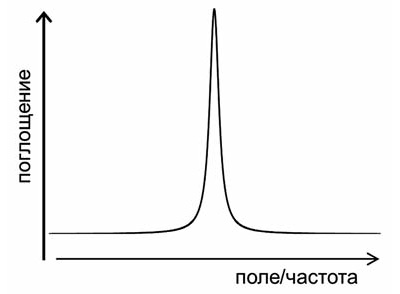
The precession frequency is determined both by the properties of the nucleus and by the strength of the magnetic field: the stronger the field, the higher the frequency. Then, if, in addition to a constant external magnetic field, an alternating magnetic field acts on the nucleus, then the nucleus begins to interact with this field - it, as it were, swings the nucleus more strongly, the precession amplitude increases, and the nucleus absorbs the energy of the alternating field. However, this will occur only under the condition of resonance, i.e., the coincidence of the precession frequency and the frequency of the external alternating field. It looks like a classic example from high school physics - soldiers marching across a bridge. If the step frequency coincides with the natural frequency of the bridge, then the bridge sways more and more. Experimentally, this phenomenon manifests itself in the dependence of the absorption of an alternating field on its frequency. At the moment of resonance, the absorption increases sharply, and the simplest magnetic resonance spectrum looks like this:
- Fourier spectroscopy
The first NMR spectrometers worked exactly as described above - the sample was placed in a constant magnetic field, and RF radiation was continuously applied to it. Then either the frequency of the alternating field or the intensity of the constant magnetic field changed smoothly. The energy absorption of the alternating field was recorded by a radio frequency bridge, the signal from which was output to a recorder or an oscilloscope. But this method of signal registration has not been used for a long time. In modern NMR spectrometers, the spectrum is recorded using pulses. The magnetic moments of the nuclei are excited by a short powerful pulse, after which a signal is recorded, which is induced in the RF coil by freely precessing magnetic moments. This signal gradually decreases to zero as the magnetic moments return to equilibrium (this process is called magnetic relaxation). The NMR spectrum is obtained from this signal using a Fourier transform. This is a standard mathematical procedure that allows you to decompose any signal into frequency harmonics and thus obtain the frequency spectrum of this signal. This method of recording the spectrum allows you to significantly reduce the noise level and conduct experiments much faster.

One excitation pulse to record the spectrum is the simplest NMR experiment. However, there can be many such pulses, of different durations, amplitudes, with different delays between them, etc., in the experiment, depending on what kind of manipulations the researcher needs to perform with the system of nuclear magnetic moments. However, almost all of these pulse sequences end in the same thing - recording a free precession signal followed by a Fourier transform.
- Magnetic interactions in matter
In itself, magnetic resonance would remain nothing more than an interesting physical phenomenon, if it were not for the magnetic interactions of nuclei with each other and with the electron shell of the molecule. These interactions affect the resonance parameters, and with their help, NMR can be used to obtain various information about the properties of molecules - their orientation, spatial structure (conformation), intermolecular interactions, chemical exchange, rotational and translational dynamics. Thanks to this, NMR has become a very powerful tool for studying substances at the molecular level, which is widely used not only in physics, but mainly in chemistry and molecular biology. An example of one of these interactions is the so-called chemical shift. Its essence is as follows: the electron shell of the molecule responds to an external magnetic field and tries to screen it - partial screening of the magnetic field occurs in all diamagnetic substances. This means that the magnetic field in the molecule will differ from the external magnetic field by a very small amount, which is called the chemical shift. However, the properties of the electron shell in different parts of the molecule are different, and the chemical shift is also different. Accordingly, the resonance conditions for nuclei in different parts of the molecule will also differ. This makes it possible to distinguish chemically nonequivalent nuclei in the spectrum. For example, if we take the spectrum of hydrogen nuclei (protons) of pure water, then there will be only one line in it, since both protons in the H 2 O molecule are exactly the same. But for methyl alcohol CH 3 OH, there will already be two lines in the spectrum (if we neglect other magnetic interactions), since there are two types of protons - protons of the methyl group CH 3 and a proton associated with an oxygen atom. As the molecules become more complex, the number of lines will increase, and if we take such a large and complex molecule as a protein, then in this case the spectrum will look something like this:

- Magnetic cores
NMR can be observed on different nuclei, but it must be said that not all nuclei have a magnetic moment. It often happens that some isotopes have a magnetic moment, while other isotopes of the same nucleus do not. In total, there are more than a hundred isotopes of various chemical elements that have magnetic nuclei, but no more than 1520 magnetic nuclei are usually used in research, everything else is exotic. Each nucleus has its own characteristic ratio of the magnetic field and the precession frequency, called the gyromagnetic ratio. For all nuclei these ratios are known. Using them, one can choose the frequency at which, for a given magnetic field, a signal from the nuclei needed by the researcher will be observed.
The most important nuclei for NMR are protons. They are most abundant in nature, and they have a very high sensitivity. For chemistry and biology, the nuclei of carbon, nitrogen and oxygen are very important, but scientists were not very lucky with them: the most common isotopes of carbon and oxygen, 12 C and 16 O, do not have a magnetic moment, the natural nitrogen isotope 14 N has a moment, but it for a number of reasons it is very inconvenient for experiments. There are 13 C, 15 N and 17 O isotopes that are suitable for NMR experiments, but their natural abundance is very low and the sensitivity is very low compared to protons. Therefore, special isotopically enriched samples are often prepared for NMR studies, in which the natural isotope of one or another nucleus is replaced by the one needed for experiments. In most cases, this procedure is very difficult and expensive, but sometimes it is the only way to get the necessary information.
- Electron paramagnetic and quadrupole resonance
Speaking of NMR, one cannot fail to mention two other related physical phenomena - electron paramagnetic resonance (EPR) and nuclear quadrupole resonance (NQR). EPR is essentially similar to NMR, the difference lies in the fact that the resonance is observed on the magnetic moments not of atomic nuclei, but of the electron shell of the atom. EPR can be observed only in those molecules or chemical groups whose electron shell contains the so-called unpaired electron, then the shell has a non-zero magnetic moment. Such substances are called paramagnets. EPR, like NMR, is also used to study various structural and dynamic properties of substances at the molecular level, but its scope is much narrower. This is mainly due to the fact that most molecules, especially in living nature, do not contain unpaired electrons. In some cases, it is possible to use a so-called paramagnetic probe, i.e. a chemical group with an unpaired electron that binds to the molecule under study. But this approach has obvious drawbacks that limit the possibilities of this method. In addition, in EPR there is no such high spectral resolution (ie, the ability to distinguish one line from another in the spectrum) as in NMR.
It is most difficult to explain the nature of NQR "on the fingers". Some nuclei have a so-called electric quadrupole moment. This moment characterizes the deviation of the distribution of the electric charge of the nucleus from spherical symmetry. The interaction of this moment with the gradient of the electric field created by the crystalline structure of the substance leads to the splitting of the energy levels of the nucleus. In this case, resonance can be observed at a frequency corresponding to transitions between these levels. Unlike NMR and EPR, NQR does not require an external magnetic field, since level splitting occurs without it. NQR is also used to study substances, but its scope is even narrower than that of EPR.
- Advantages and disadvantages of NMR

NMR is the most powerful and informative method for studying molecules. Strictly speaking, this is not one method, but a large number of different types of experiments, i.e., pulse sequences. Although they are all based on the NMR phenomenon, but each of these experiments is designed to obtain some specific specific information. The number of these experiments is measured by many tens, if not hundreds. Theoretically, NMR can, if not everything, then almost everything that all other experimental methods for studying the structure and dynamics of molecules can, although in practice this is, of course, far from always feasible. One of the main advantages of NMR is that, on the one hand, its natural probes, i.e., magnetic nuclei, are distributed over the entire molecule, and, on the other hand, it makes it possible to distinguish these nuclei from each other and obtain spatially selective data on properties of the molecule. Almost all other methods provide information either averaged over the entire molecule, or only about one of its parts.
There are two main disadvantages of NMR. First, this is a low sensitivity compared to most other experimental methods (optical spectroscopy, fluorescence, EPR, etc.). This leads to the fact that in order to average the noise, the signal must be accumulated for a long time. In some cases, the NMR experiment can be carried out for even several weeks. Secondly, it is its high cost. NMR spectrometers are among the most expensive scientific instruments, costing at least hundreds of thousands of dollars, with the most expensive spectrometers costing several million. Not all laboratories, especially in Russia, can afford to have such scientific equipment.
- Magnets for NMR spectrometers
One of the most important and expensive parts of a spectrometer is the magnet, which creates a constant magnetic field. The stronger the field, the higher the sensitivity and spectral resolution, so scientists and engineers are constantly trying to get the highest possible fields. The magnetic field is created by an electric current in the solenoid - the stronger the current, the greater the field. However, it is impossible to increase the current indefinitely; at a very high current, the solenoid wire will simply begin to melt. Therefore, superconducting magnets, i.e., magnets in which the solenoid wire is in the superconducting state, have been used for a very long time for high-field NMR spectrometers. In this case, the electrical resistance of the wire is zero, and no energy is released at any current value. The superconducting state can only be obtained at very low temperatures, just a few degrees Kelvin - this is the temperature of liquid helium. (High-temperature superconductivity is still only a matter of purely fundamental research.) It is with the maintenance of such a low temperature that all the technical difficulties in the design and production of magnets are connected, which cause their high cost. The superconducting magnet is built on the principle of a thermos matryoshka. The solenoid is in the center, in the vacuum chamber. It is surrounded by a shell containing liquid helium. This shell is surrounded by a shell of liquid nitrogen through a vacuum layer. The temperature of liquid nitrogen is minus 196 degrees Celsius, nitrogen is needed so that helium evaporates as slowly as possible. Finally, the nitrogen shell is isolated from room temperature by an outer vacuum layer. Such a system is able to maintain the desired temperature of the superconducting magnet for a very long time, although this requires regular pouring of liquid nitrogen and helium into the magnet. The advantage of such magnets, in addition to the ability to obtain high magnetic fields, is also that they do not consume energy: after the start of the magnet, the current runs through the superconducting wires with virtually no loss for many years.
- Tomography
In conventional NMR spectrometers, they try to make the magnetic field as uniform as possible, this is necessary to improve the spectral resolution. But if the magnetic field inside the sample, on the contrary, is made very inhomogeneous, this opens up fundamentally new possibilities for using NMR. The inhomogeneity of the field is created by the so-called gradient coils, which are paired with the main magnet. In this case, the magnitude of the magnetic field in different parts of the sample will be different, which means that the NMR signal can be observed not from the entire sample, as in a conventional spectrometer, but only from its narrow layer, for which the resonance conditions are met, i.e., the desired ratio of magnetic field and frequency. By changing the magnitude of the magnetic field (or, which is essentially the same thing, the frequency of observing the signal), you can change the layer that will give the signal. Thus, it is possible to "scan" the sample throughout its volume and "see" its internal three-dimensional structure without destroying the sample in any mechanical way. To date, a large number of techniques have been developed that make it possible to measure various NMR parameters (spectral characteristics, magnetic relaxation times, self-diffusion rate, and some others) with spatial resolution inside a sample. The most interesting and important, from a practical point of view, the use of NMR tomography was found in medicine. In this case, the "sample" being examined is the human body. NMR imaging is one of the most effective and safe (but also expensive) diagnostic tools in various fields of medicine, from oncology to obstetrics. It is curious to note that doctors do not use the word "nuclear" in the name of this method, because some patients associate it with nuclear reactions and the atomic bomb.
- Discovery history

The year of the discovery of NMR is considered to be 1945, when the Americans Felix Bloch from Stanford and independently Edward Parcell and Robert Pound from Harvard first observed the NMR signal on protons. By that time, much was already known about the nature of nuclear magnetism, the NMR effect itself was theoretically predicted, and several attempts were made to observe it experimentally. It is important to note that a year earlier in the Soviet Union, in Kazan, the EPR phenomenon was discovered by Evgeny Zavoisky. It is now well known that Zavoisky also observed the NMR signal, this was before the war, in 1941. However, he had a poor quality magnet with poor field uniformity at his disposal, the results were poorly reproducible and therefore remained unpublished. In fairness, it should be noted that Zavoisky was not the only one who observed NMR before its "official" discovery. In particular, the American physicist Isidore Rabi (Nobel Prize winner in 1944 for the study of the magnetic properties of nuclei in atomic and molecular beams) also observed NMR in the late 1930s, but considered this to be an instrumental artifact. One way or another, but our country remains a priority in the experimental detection of magnetic resonance. Although Zavoisky himself soon after the war began to deal with other problems, his discovery for the development of science in Kazan played a huge role. Kazan is still one of the world's leading research centers for EPR spectroscopy.
- Nobel Prizes in Magnetic Resonance
In the first half of the 20th century, several Nobel Prizes were awarded to scientists without whose work the discovery of NMR could not have taken place. Among them are Peter Szeeman, Otto Stern, Isidor Rabi, Wolfgang Pauli. But there were four Nobel Prizes directly related to NMR. In 1952, Felix Bloch and Edward Purcell received the prize for the discovery of NMR. This is the only "NMR" Nobel Prize in physics. In 1991, the Swiss Richard Ernst, who worked at the famous ETH Zurich, won the Chemistry Prize. He was awarded it for the development of multidimensional NMR spectroscopy methods, which made it possible to radically increase the information content of NMR experiments. In 2002, the prize winner, also in chemistry, was Kurt Wüthrich, who worked with Ernst in neighboring buildings at the same Technical School. He received the award for developing methods for determining the three-dimensional structure of proteins in solution. Prior to this, the only method that allowed determining the spatial conformation of large biomacromolecules was only X-ray diffraction analysis. Finally, in 2003, the American Paul Lauterbur and the Englishman Peter Mansfield received the Medical Prize for the invention of NMR imaging. The Soviet discoverer of the EPR E.K. Zavoisky, alas, did not receive the Nobel Prize.
MINISTRY OF HEALTH OF THE RUSSIAN FEDERATION
GENERAL PHARMACOPEIAN AUTHORIZATION
Spectroscopy of nuclear GPM.1.2.1.1.0007.15
magnetic resonance instead of GFXII, part 1,
OFS 42-0046-07
Nuclear magnetic resonance spectroscopy (NMR) is a method based on the absorption of radio frequency electromagnetic radiation by the nuclei of a sample with a nonzero magnetic moment placed in a constant magnetic field ( B 0). Non-zero magnetic moments have isotopes of nuclei of elements with an odd atomic mass (1 H, 13 C, 15 N, 19 F, 31 P, etc.).
General principles
A nucleus rotating around its axis has its own moment of momentum (angular momentum, or spin) P. The magnetic moment of the nucleus μ is directly proportional to the spin: μ = γ ∙ P(γ is the proportionality factor or gyromagnetic ratio). The angular and magnetic moments are quantized, i.e. can be in one of 2 I+ 1 spin states ( I – spin quantum number). Different states of the magnetic moments of nuclei have the same energy if they are not affected by an external magnetic field. When nuclei are placed in an external magnetic field B 0, the energy degeneracy of the nuclei is removed and the possibility of an energy transition from one level to another arises. The process of distribution of nuclei between different energy levels proceeds in accordance with the Boltzmann distribution law and leads to the appearance of a macroscopic equilibrium longitudinal magnetization M z . The time it takes to create M z after turning on the external magnetic field AT 0 , is called time longitudinal or spin—lattice relaxation (T one). Violation of the equilibrium distribution of nuclei occurs under the action of a radio frequency magnetic field ( B 1), perpendicular B 0 , which causes additional transitions between energy levels, accompanied by energy absorption (the phenomenon nuclear magnetic resonance). Frequency ν 0 , at which the absorption of energy by nuclei occurs ( Larmorova or resonant absorption frequency), varies depending on the value of the constant field B 0: ν 0 = γ B 0 /2π. At the moment of resonance, there is an interaction between the individual nuclear magnetic moments and the field AT 1 , which outputs a vector M z from its equilibrium position along the axis z. As a result, there appears transverse magnetization M xy. Its change associated with the exchange within the spin system is characterized by the time transverse or spin-spin relaxation (T 2).
Dependence of the intensity of energy absorption by nuclei of the same type on the frequency of the radio-frequency magnetic field at a fixed value AT 0 is called one-dimensional spectrumnuclear magnetic resonance kernels of this type. An NMR spectrum can be obtained in two ways: by continuously irradiating the sample with an RF field of varying frequency, as a result of which the NMR spectrum is recorded directly (continuous exposure spectroscopy), or by exposing the sample to a short RF pulse ( pulsed spectroscopy). In pulsed NMR spectroscopy, time-decayed coherent radiation emitted by nuclei upon returning to the initial spin state ( free induction decay signal) followed by the transformation of the time scale into frequency ( Fourier transform).
In molecules, the electrons of atoms reduce the magnitude of the acting external magnetic field B 0 at the location of the kernel, i.e. appears diamagnetic shielding:
B loc = B 0 ∙ (1 – σ),
B lok is the intensity of the resulting field;
σ is the screening constant.
The difference in the resonant frequencies of the signals of the nuclei, equal to the difference in their screening constants, is called chemical shift signals, indicated by the symbol δ , measured in parts per million (ppm). Interaction of magnetic moments of nuclei through chemical bond electrons ( spin-spin interaction) causes splitting of the NMR signal ( multiplicity, m). The number of components in multiplets is determined by the nuclear spin and the number of interacting nuclei. The measure of the spin-spin interaction is spin-spin coupling constant (J, measured in hertz, Hz). Values δ, m and J do not depend on the magnitude of the constant magnetic field.
The intensity of the nuclear NMR signal in the spectrum is determined by the population of its energy levels. Of the nuclei with a natural abundance of isotopes, the most intense signals are produced by hydrogen nuclei. The intensity of NMR signals is also affected by the time of longitudinal-transverse relaxation (large T 1 lead to a decrease in signal intensity).
The width of NMR signals (difference between frequencies at half maximum of the signal) depends on T 1 and T 2. small times T 1 and T 2 cause wide and poorly interpreted spectrum signals.
The sensitivity of the NMR method (maximum detectable concentration of a substance) depends on the intensity of the nuclear signal. For 1 H nuclei, the sensitivity is 10 -9 ÷ 10 -11 mol.
Correlations of various spectral parameters (for example, chemical shifts of different nuclei within the same molecular system) can be obtained by homo- and heteronuclear methods in 2D or 3D format.
device
High resolution NMR pulse spectrometer (NMR spectrometer) consists of:
- magnet to create a constant magnetic field B 0 ;
- a temperature-controlled sensor with a sample holder for applying an RF pulse and detecting the radiation emitted by the sample;
- an electronic device for creating a radio frequency pulse, recording, amplifying and converting the free induction decay signal into digital form;
- devices for tuning and adjusting electronic circuits;
- data collection and processing devices (computer);
and may also include:
a flow cell for NMR liquid chromatography or flow-injection analysis;
- system for creating a pulsed magnetic field gradient.
A strong magnetic field is generated by a superconductivity coil in a Dewar vessel filled with liquid helium.
The proper functioning of the NMR spectrometer should be checked. For verification, appropriate tests are carried out, including, as a rule, the measurement of the spectral linewidth at half-height of certain peaks under certain conditions ( permission), signal position reproducibility and signal-to-noise ratio (the ratio between the intensity of a specific signal in the NMR spectrum and random fluctuations in the region of the spectrum that does not contain signals from the analyte, S/N) for standard mixtures. The spectrometer software contains algorithms for determining S/N. All instrument manufacturers provide specifications and measurement protocols for these parameters.
NMR Spectroscopy of Samples in Solutions
Methodology
The test sample is dissolved in a solvent to which an appropriate chemical shift calibration standard may be added as specified in the regulatory document. The value of the relative chemical shift of the nucleus of a substance (δ in-in) is determined by the following expression:
δ in-in \u003d (ν in-in - ν standard) / ν of the device,
ν in-in - the resonance frequency of the core of the substance, Hz;
ν etalon is the resonance frequency of the etalon core, Hz;
ν of the device is the operating frequency of the NMR spectrometer (the frequency at which the resonance conditions for hydrogen nuclei are satisfied for a given B 0, MHz).
For solutions in organic solvents, the chemical shift in the 1H and 13C spectra is measured relative to the tetramethylsilane signal, the position of which is taken as 0 ppm. The chemical shifts are counted in the direction of a weak field (to the left) from the tetramethylsilane signal (delta is the scale of chemical shifts). For aqueous solutions, sodium 2,2-dimethyl-2-silanepentane-5-sulfonate is used as a reference in the 1 H NMR spectra, the chemical shift of the protons of the methyl group of which is 0.015 ppm. For the spectra of 13 C aqueous solutions, dioxane is used as a reference, the chemical shift of which is 67.4 ppm.
When calibrating the 19 F spectra, trifluoroacetic acid or trichlorofluoromethane is used as the primary standard with zero chemical shift; spectra 31 P - 85% solution of phosphoric acid or trimethyl phosphate; spectra 15 N - nitromethane or saturated ammonia solution. In 1 H and 13 C NMR, as a rule, an internal standard is used, which is directly added to the test sample. 15 N, 19 F, and 31 P NMR often use an external standard, which is held separately in a coaxial cylindrical tube or capillary.
When describing NMR spectra, it is necessary to indicate the solvent in which the substance is dissolved and its concentration. Easily mobile liquids are used as solvents, in which hydrogen atoms are replaced by deuterium atoms to reduce the intensity of solvent signals. The deuterated solvent is selected based on the following criteria:
- 1) the solubility of the test compound in it;
- 2) no overlap between the signals of residual protons of the deuterated solvent and the signals of the test compound;
- 3) no interaction between the solvent and the test compound, unless otherwise indicated.
Solvent atoms give signals that are easily identified by their chemical shift and can be used to calibrate the chemical shift axis (secondary standard). The chemical shifts of the residual proton signals of deuterated solvents have the following values (ppm): chloroform, 7.26; benzene, 7.16; water - 4.7; methanol -3.35 and 4.78; dimethyl sulfoxide - 2.50; acetone - 2.05; the position of the signal of water and the protons of the hydroxyl groups of alcohols depends on the pH of the medium and temperature.
For quantitative analysis, solutions must be free of undissolved particles. For some assays, it may be necessary to add an internal standard to compare test and reference intensities. Appropriate standard samples and their concentrations should be specified in the normative documentation. After placing the sample in a test tube and capping, the sample is introduced into the magnet of the NMR spectrometer, the test parameters are set (settings, registration, digitization of the free induction decay signal). The main test parameters given in the regulatory documentation are recorded or stored in a computer.
To prevent spectrum drift over time, a stabilization procedure (deuterium lock) is performed using the deuterium signal induced by deuterated solvents, unless otherwise indicated. The instrument is adjusted to obtain the most optimal resonance conditions and the maximum ratio S/N(shimming).
During the test, it is possible to perform multiple sequences of cycles "impulse - data acquisition - pause" with subsequent summation of individual signals of the decay of free induction and averaging the noise level. The delay time between pulse sequences during which the system of nuclear spins restores its magnetization ( D 1), for quantitative measurements must exceed the longitudinal relaxation time T 1: D 1 ≥ 5 T one . The spectrometer software contains algorithms for determining T one . If the value T 1 is unknown, it is recommended to use the value D 1 = 25 sec.
After carrying out the Fourier transform, the signals in the frequency representation are calibrated to the selected standard and their relative intensity is measured by integration - measuring the ratio of the areas of the resonant signals. In the 13 C spectra, only signals of the same type are integrated. The signal integration accuracy depends on the ratio signal – noise (S/N):
where u(I) is the standard uncertainty of integration.
The number of free induction decay accumulations required to achieve a satisfactory ratio S/ N, should be given in the regulatory documentation.
Along with one-dimensional for analytical purposes, homo- and heteronuclear two-dimensional correlation spectra are used, based on a certain sequence of pulses (COSY, NOESY, ROESY, HSQC, HMBC, HETCOR, CIGAR, INADEQUATE, etc.). In two-dimensional spectra, the interaction between nuclei manifests itself in the form of signals called cross peaks. The position of the cross peaks is determined by the values of the chemical shifts of the two interacting nuclei. Two-dimensional spectra are preferably used to determine the composition of complex mixtures and extracts, because the probability of signal superposition (cross peaks) in two-dimensional spectra is significantly lower than the probability of signal superposition in one-dimensional spectra.
To quickly obtain the spectra of heteronuclei (13 C, 15 N, etc.), methods (HSQC, HMBC) are used, which make it possible to obtain spectra of other nuclei on 1 H nuclei using the mechanisms of heteronuclear interaction.
The DOSY technique, based on recording the loss of phase coherence of nuclear spins due to translational displacements of molecules under the action of a magnetic field gradient, makes it possible to obtain spectra of individual compounds (spectral separation) in a mixture without their physical separation and to determine the sizes, degrees of aggregation, and molecular weights of molecular objects (molecules , macromolecules, molecular complexes, supramolecular systems).
Areas of use
The variety of structural and analytical information contained in nuclear magnetic resonance spectra makes it possible to use the nuclear magnetic resonance method for qualitative and quantitative analysis. The use of nuclear magnetic resonance spectroscopy in quantitative analysis is based on the direct proportionality of the molar concentration of magnetically active nuclei to the integrated intensity of the corresponding absorption signal in the spectrum.
- Identification of the active substance. The identification of the active substance is carried out by comparing the spectrum of the test sample with the spectrum of a standard sample or with a published reference spectrum. The spectra of standard and test samples should be obtained using the same methods and conditions. The peaks in the compared spectra should coincide in position (deviations of the values δ test and standard samples within ± 0.1 ppm. for nuclear magnetic resonance 1 N and ± 0.5 ppm. for nuclear magnetic resonance 13 C), integrated intensity and multiplicity, the values of which should be given when describing the spectra. In the absence of a standard sample, a pharmacopoeial standard sample can be used, the identity of which is confirmed by independent structural interpretation of the spectral data and alternative methods.
When confirming the authenticity of samples of non-stoichiometric composition (for example, natural polymers of variable composition), the peaks of the test and standard samples are allowed to differ in position and integral intensity of the signals. The spectra to be compared must be similar, i.e. contain the same characteristic regions of the signals, confirming the coincidence of the fragment composition of the test and standard samples.
To establish the authenticity of a mixture of substances (extracts), one-dimensional NMR spectra can be used as a whole, as “fingerprints” of an object, without detailing the values of δ and the multiplicity of individual signals. In the case of using two-dimensional NMR spectroscopy in the description of spectra (spectrum fragments) claimed for authenticity, the values of cross peaks should be given.
- Identification of foreign matter/residual organic solvents. Identification of impurities/residual organic solvents is carried out similarly to the identification of the active substance, tightening the requirements for sensitivity and digital resolution.
- Determination of the content of foreign impurities / residual organic solvents in relation to the active substance. The NMR method is a direct absolute method for determining the molar ratio of the active substance and the impurity compound ( n/n impurity):

where S‘ and S‘ impurity - normalized values of the integral intensities of the signals of the active substance and impurity.
Normalization is carried out according to the number of nuclei in the structural fragment, which determine the measured signal.
Mass fraction of impurity / residual organic solvent relative to the active substance ( X pr) is determined by the formula:

M pr is the molecular weight of the impurity;
M is the molecular weight of the active substance;
S‘ pr is the normalized value of the integral intensity of the impurity signal;
S'– normalized value of the integral intensity of the signal of the active substance.
- Quantitative determination of the content of the substance (active substance, impurity / residual solvent) in the pharmaceutical substance. Absolute content of matter in a pharmaceutical substance, it is determined by the internal standard method, which is chosen as a substance whose signals are close to the signals of the analyte, without overlapping with them. The signal intensities of the analyte and the standard should not differ significantly.
The percentage of the analyte in the test sample in terms of dry matter ( x,% mass) is calculated by the formula:
x,% mass = 100 ∙ ( S‘ /S‘ 0) ∙ (M ∙ a 0 /M 0 ∙ a) ∙ ,
S' is the normalized value of the integral intensity of the signal of the analyte;
S‘ 0 is the normalized value of the integrated signal intensity of the standard;
M is the molecular weight of the analyte;
M 0 – molecular weight;
a- weighing of the test sample;
a 0– weight of the standard substance;
W- moisture contents, %.
The following compounds can be used as standards: maleic acid (2H; 6.60 ppm, M= 116.07), benzyl benzoate (2H; 5.30 ppm, M= 212.25), malonic acid (2H; 3.30 ppm, M= 104.03), succinimide (4H; 2.77 ppm, M= 99.09), acetanilide (3H; 2.12 ppm, M = 135,16), tert-butanol (9H; 1.30 ppm, M = 74,12).
Relative substance content as the proportion of a component in a mixture of components of a pharmaceutical substance is determined by the method of internal normalization. molar ( X mol) and mass ( X mass) component fraction i in a mixture n substances is determined by the formulas:

- Determination of the molecular weight of proteins and polymers. The molecular weights of proteins and polymers are determined by comparing their mobility with that of reference compounds of known molecular weight using DOSY techniques. Self-diffusion coefficients are measured ( D) of the test and standard samples, build a graph of the dependence of the logarithms of the molecular weights of the standard compounds on the logarithms D. From the graph thus obtained, the unknown molecular weights of the test samples are determined by linear regression. A full description of the DOSY experiment should be given in the regulatory documentation.
NMR spectroscopy of solids
Samples in the solid state are analyzed using specially equipped NMR spectrometers. Certain technical operations (rotation of a powdered sample in a rotor inclined at a magic angle (54.7°) to the magnetic field axis AT 0 , force depairing, polarization transfer from highly excitable nuclei to less polarizable nuclei - cross-polarization) make it possible to obtain high-resolution spectra of organic and inorganic compounds. A full description of the procedure should be given in the regulatory documentation. The main area of application of this type of NMR spectroscopy is the study of polymorphism of solid drugs.
Today, more and more patients are referred not for radiography or ultrasound, but for nuclear magnetic resonance imaging. This research method is based on core magnetism. Let's consider what NMR tomography is, what are its advantages and in what cases it is performed.
What is this study?
This diagnostic method is based on nuclear magnetic resonance. In an external magnetic field, the nucleus of a hydrogen atom, or proton, is in two mutually opposite states. You can change the direction of the magnetic moment of the nucleus by acting on it with electromagnetic rays with a certain frequency.
Placing a proton in an external magnetic field causes a change in its magnetic moment with a return to its original position. This releases a certain amount of energy. Magnetic resonance tomography captures the change in the amount of such energy.
The tomograph uses very strong magnetic fields. Electromagnets are usually capable of developing a magnetic field with a strength of 3, sometimes up to 9 T. It is completely harmless to humans. The tomography system allows you to localize the direction of the magnetic field in order to obtain the highest quality images.
Nuclear magnetic tomograph
The diagnostic method is based on fixing the electromagnetic response of the nucleus of an atom (proton), which occurs due to its excitation by electromagnetic waves in a high-voltage magnetic field. Magnetic resonance imaging was first discussed in 1973. Then the American scientist P. Laterbur proposed to study the object in a changing magnetic field. The works of this scientist served as the beginning of a new era in medicine.
With the help of a magnetic resonance tomograph, it became possible to study the tissues and cavities of the human body due to the degree of tissue saturation with hydrogen. Magnetic resonance imaging contrast agents are often used. Most often, these are preparations of gadolinium, which are able to change the response of protons.
The term "nuclear MRI" existed until 1986.
In connection with radiophobia among the population in connection with the disaster at the Chernobyl nuclear power plant, it was decided to remove the word “nuclear” from the name of the new diagnostic method. However, this allowed magnetic resonance imaging to quickly enter the practice of diagnosing many diseases. Today, this method is the key to identifying many more recently difficult-to-diagnose diseases.
How is the diagnosis carried out?
An MRI uses a very strong magnetic field. And although it is not dangerous for humans, nevertheless, the doctor and the patient need to adhere to certain rules.
First of all, before the diagnostic procedure, the patient fills out a special questionnaire. In it, he indicates the state of health, as well as statements about himself. The examination is done in a specially prepared room with a cabin for changing clothes and personal belongings.
In order not to harm himself, and also to ensure the correctness of the results, the patient should take off all things that contain metal, leave mobile phones, credit cards, watches, etc. in the locker for personal belongings. It is desirable for women to wash off decorative cosmetics from the skin.
Next, the patient is placed inside the tomograph tube. At the direction of the doctor, the examination area is determined. Each zone is examined for ten to twenty minutes. During this time, the patient must remain still. The quality of the pictures will depend on this. The doctor can fix the position of the patient, if necessary.
During the operation of the device, uniform sounds are heard. This is normal and indicates that the study is proceeding correctly. To obtain more accurate results, a contrast agent may be administered intravenously to the patient. In some cases, with the introduction of such a substance, a surge of heat is felt. This is completely normal.
Approximately half an hour after the study, the doctor can receive the study protocol (conclusion). A disk with the results is also issued.
Benefits of Nuclear MRI
The benefits of such a survey include the following.
- The ability to obtain high-quality images of body tissues in three projections. This greatly enhances the visualization of tissues and organs. In this case, MRI is much better than computed tomography, radiography and ultrasound diagnostics.
- High-quality 3D images provide an accurate diagnosis, which improves treatment and increases the likelihood of recovery.
- Since it is possible to obtain a high-quality image on an MRI, such a study is the best for detecting tumors, disorders of the central nervous system, and pathological conditions of the musculoskeletal system. Thus, it becomes possible to diagnose those diseases that until recently were difficult or impossible to detect.
- Modern devices for tomography allow you to get high-quality images without changing the position of the patient. And for encoding information, the same methods are used as in computed tomography. This facilitates diagnosis, as the doctor sees three-dimensional images of entire organs. Also, the doctor can get images of a particular organ in layers.
- Such an examination well determines the earliest pathological changes in the organs. Thus, it is possible to detect the disease at a stage when the patient does not yet feel symptoms.
- During such a study, the patient is not exposed to ionizing radiation. This significantly expands the scope of MRI.
- The MRI procedure is completely painless and does not cause any discomfort to the patient.
Indications for MRI
There are many indications for magnetic resonance imaging.
- Cerebral circulation disorders.
- Suspicions of a neoplasm of the brain, damage to its membranes.
- Assessment of the state of organs after surgery.
- Diagnosis of inflammatory phenomena.
- Convulsions, epilepsy.
- Traumatic brain injury.
- Assessment of the condition of the vessels.
- Assessment of the condition of bones and joints.
- Diagnosis of the soft tissues of the body.
- Diseases of the spine (including osteochondrosis, spondyloarthrosis).
- Spinal injury.
- Assessment of the state of the spinal cord, including suspicion of malignant processes.
- Osteoporosis.
- Assessment of the state of the peritoneal organs, as well as the retroperitoneal space. MRI is indicated for jaundice, chronic hepatitis, cholecystitis, cholelithiasis, tumor-like liver damage, pancreatitis, diseases of the stomach, intestines, spleen, kidneys.
- Diagnosis of cysts.
- Diagnosis of the state of the adrenal glands.
- Diseases of the pelvic organs.
- Urological pathologies.
- Gynecological diseases.
- Diseases of the organs of the chest cavity.
In addition, magnetic resonance imaging of the whole body is indicated if a neoplasm is suspected. MRI can be used to search for metastases if a primary tumor is diagnosed.
This is not a complete list of indications for magnetic resonance imaging. It is safe to say that there is no such organism and disease that could not be detected using this diagnostic method. Since the possibilities of medicine are growing, doctors have practically unlimited possibilities for diagnosing and treating many dangerous diseases.
When is magnetic resonance imaging contraindicated?
There are a number of absolute and relative contraindications for MRI. Absolute contraindications include:
- Presence of a pacemaker. This is due to the fact that fluctuations in the magnetic field are able to adapt to the rhythm of the heart and thus can be fatal.
- The presence of installed ferromagnetic or electronic implants in the middle ear.
- Large metal implants.
- The presence of ferromagnetic fragments in the body.
- Availability of Ilizarov apparatus.
Relative contraindications (when research is possible under certain conditions) include:

When performing MRI with contrast, contraindications are anemia, chronic decompensated renal failure, pregnancy, individual intolerance.
Conclusion
The importance of magnetic resonance imaging for diagnosis cannot be overestimated. It is a perfect, non-invasive, painless and harmless way of detecting many diseases. With the introduction of magnetic resonance imaging, the treatment of patients has also improved, as the doctor knows accurate diagnosis and features of all processes occurring in the patient's body.
No need to be afraid of an MRI. The patient does not feel any pain during the procedure. It has nothing to do with nuclear or x-ray radiation. It is also impossible to refuse such a procedure.
